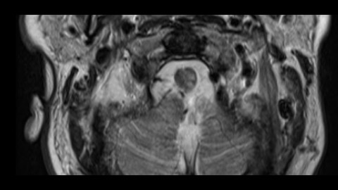
Spinal cord lesions
A collection of spinal cord imaging cases with emphasis on demyelinating, infectious, and inflammatory conditions with a few tumors for contrast. More cases added weekly so check back soon!
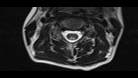
Multiple Sclerosis - Spinal Cord
Multiple typical spinal cord lesions of Multiple Sclerosis. They are pretty focal and do not span more than 3 vertebral levels.
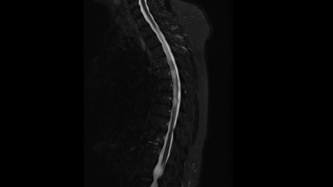
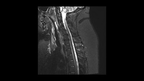
Multiple Sclerosis - Thoracic Cord
Multiple cord lesions of Multiple Sclerosis in the thoracic spine.
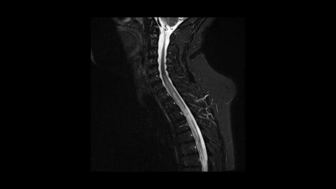
Neuromyelitis optica
A case of neuromyelitis optica with avid enhancement.
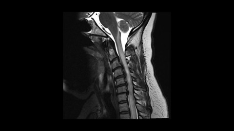
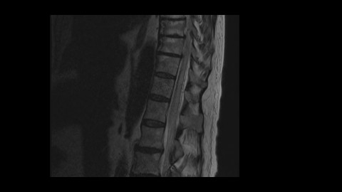
Spondylotic Myelopathy with enhancement
This is a case of spondylotic myelopathy presenting with longitudinally extensive myelopathy extending both above and below the levels of spinal cord compression as well as pronounced enhancement. Post decompression images show marked improvement of the T2 hyperintensity and resolution of the enhancement. Keep this pattern of spondylotic myelopathy in mind as it can be mistaken for demyelinating diseases or even tumors.
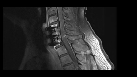
Spondylotic Myelopathy with enhancement
Another case of spondylotic myelopathy with enhancement. Go through the keyfindings in order for some more discussion (@Key Finding 1, @Key Finding 2, @Key Finding 3, @Key Finding 4, @Key Finding 5).
Cobalamin deficiency - Subacute combined degeneration
Typical symmetric dorsal column T2 hyperintensity seen with subacute combined degeneration due to vitamin B12 deficiency.
Spinal dural arteriovenous fistula
A case of dural arteriovenous fistula. Notice prominent signal voids (@Key Finding 2). Angiographic images demonstrated arterial feeding from the ascending pharyngeal branch of the external carotid artery (@Key Finding 3)
Spinal dural arteriovenous fistula
A case of spinal dural arteriovenous fistula (@Key Finding 1, @Key Finding 2)
Upper cord infarct
A case of upper spinal cord, cerviomedullary junction infarctions (@Key Finding 2, @Key Finding 3)